- Research note
- Open access
- Published:
Cytokeratin 5/6 expression in bladder cancer: association with clinicopathologic parameters and prognosis
BMC Research Notes volume 11, Article number: 207 (2018)
Abstract
Objectives
Well differentiated keratinized squamous component as a part of urothelial carcinoma can be easily appreciated; however non-keratinizing squamous differentiation closely resembles urothelial differentiation. In addition prognostic significance of CK 5/6 expression in the absence of apparent squamous differentiation is still unclear. Therefore, in the present study we aimed to evaluate the frequency of CK 5/6 expression in 127 cases of urothelial carcinoma and its prognostic significance in loco-regional population.
Results
Positive CK5/6 expression was noted in 6.3% (8 cases) and 13.4% (17 cases) revealed focal positive CK 5/6 expression. On the other hand, 80.3% (102 cases) showed negative CK5/6 staining. Significant association of CK5/6 expression was noted with tumor grade and muscularis propria invasion, however no significant association was noted with overall and disease free survival. On the basis of the results of our study we can conclude that CK5/6 is an independent prognostic biomarker in urothelial carcinoma and therefore can be used in the prognostic stratification of the patients with bladder cancer.
Introduction
Bladder cancer is among one of the most common malignancy in males worldwide and its incidence is even higher in developing countries owing to certain endemic infections [1, 2]. Urothelial carcinoma is the most common histologic subtype of bladder cancer, while Squamous cell carcinoma is seen in association with bladder stones and schistosomiasis. Muscle invasion is one of the most important prognostic factors in bladder cancer; as it necessitates radical therapy and poor 5 year disease free survival [3, 4]. While, well differentiated keratinized squamous component as part of urothelial carcinoma can be easily appreciated; non-keratinizing squamous differentiation closely resembles urothelial differentiation. On the other hand, WHO/ISUP don’t recommend routine use of immunohistochemical markers to identify squamous differentiation in urothelial carcinoma. Conversely, markers of squamous differentiation like p63, p40, CK5/6 can be positive in urothelial carcinoma [5]. In addition prognostic significance of CK 5/6 expression in the absence of apparent squamous differentiation is still unclear. Therefore, in the present study we aimed to evaluate the frequency of CK 5/6 expression in urothelial carcinoma and its prognostic significance in loco-regional population.
Main text
Total 240 diagnosed cases of urothelial carcinoma specimens were selected from records of pathology department. All patients underwent surgeries at Liaquat National hospital, Karachi from January 2010 till December 2014 over a period of 5 years. The study was approved by research and ethical review committee of Liaquat National Hospital and informed written consent was taken from all patients at the time of surgery. Hematoxylin and eosin stained slides and paraffin blocks of all cases were retrieved and new sections were cut when necessary. Slides of all cases were reviewed by two senior histopathologists and pathologic characteristics like histologic type, tumor grade, lamina propria invasion, muscularis propria invasion were evaluated. Clinical records of 61 patients were available and are thus reviewed from institutional records to evaluate history of radiation and chemotherapy and recurrence status. Moreover, representative tissue blocks of 127 cases were available for CK5/6 immunohistochemistry.
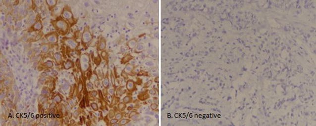
CK5/6 IHC was performed by using FLEX Monoclonal Mouse Anti-human Cytokeratin 5/6, clone D5/16 B4 (Lot No. 20042129) by DAKO envision method according to manufacturers protocol on 127 cases of urothelial carcinoma (on representative tissue blocks). Results of IHC staining were interpreted by two senior histopathologists with more than 5 years experience of reporting histopathology and immunohistochemistry and they were blinded by other histopathological features of the tumors. For quantification, at least 1000 cells were counted in 10 HPFs (40×). Intermediate to strong cytoplasmic and membranous staining in more than 10% of tumor cells was considered positive. Weak to intermediate staining in < 10% was taken as focal positive, while no staining was considered as negative (Fig. 1).
Recurrence status and follow-up were evaluated by reviewing hospital medical records. Overall survival was taken as time from surgical excision till death or last follow-up and disease free survival was defined as time between surgical excision and local recurrence or distant metastasis, death or last follow-up.
All cases of primary urothelial carcinoma were included in the study. Cases of squamous cell carcinoma or those cases of urothelial carcinoma showing divergent differentiation (including squamous differentiation) were excluded from the study.
Statistical package for social sciences (SPSS 21) was used for data compilation and analysis. Mean and standard deviation were calculated for quantitative variables. Frequency and percentage were calculated for qualitative variables. Chi square was applied to determine association. Student t test or Mann Witney test were applied to compare difference in means among groups. Survival curves were plotted using Kaplan–Meier method and the significance of difference between survival curves were determined using log-rank ratio. P value ≤ 0.05 was taken as significant.
Mean age of patients was 63.23 + 13.9 years with male to female ratio of 3:1. 95.8% specimens were of transurethral resections. 50.8% (122 cases) were of high grade morphology, whereas 49.2% (118 cases) showed low grade histology. Lamina propria invasion was seen in 30.4% (73 cases), while muscularis propria invasion was noted in 22.9% (55 cases). Mean follow up of patients involved in the study was 22.0 + 13.74 months and recurrence was seen in 45.9% (28 cases) as presented in Table 1.
Positive CK5/6 expression was noted in 6.3% (8 cases) and 13.4% (17 cases) revealed focal positive CK 5/6 expression. On the other hand, 80.3% (102 cases) showed negative CK5/6 staining. Significant association of CK5/6 expression was noted with tumor grade and muscularis propria invasion, however no significant association was noted with lamina propria invasion and disease free survival (Table 2 and Figs. 2 and 3).
In the present study we found that CK5/6 expression is low in urothelial carcinoma in our set up; however, its positivity signifies adverse prognostic features like higher tumor grade and muscularis propria invasion.
CK5/6 is a basal cytokeratin which normally expresses in squamous epithelium and in squamous cell carcinoma. Although diagnosis of squamous cell carcinoma in bladder is restricted to those tumors which show pure squamous differentiation in the absence of any urothelial component. Conversely, advanced urothelial carcinoma can show divergent differentiation (including squamous component) in up to 50% of cases and is associated with poor disease progression [6]. Morphologic diagnosis of squamous differentiation in urothelial carcinoma is based on the presence of either intercellular bridges or presence of keratinization in the form of keratin pearls or individual cell keratinization; however non-keratinizing or poorly differentiated squamous component can closely resemble urothelial carcinoma and therefore can’t be readily apparent. Gaisa et al. [7] performed IHC markers of squamous differentiation including CK5/6 and CK4/14; and found squamous differentiation in a high proportion of urothelial carcinoma without morphologic evidence of squamous differentiation. Langer et al. evaluated the prognostic value of keratin subtyping in urothelial carcinoma and revealed the prognostic impact of various cytokeratin staining in urothelial carcinoma including CK5/6.
Limitations
One of the major limitations of our study was that we performed only single biomarker of squamous differentiation in our study; use of multiple markers like CK5/14 and CK4/14 could increase the sensitivity of the study. However, on the basis of the results of our study we can conclude that CK5/6 is an independent prognostic biomarker in urothelial carcinoma and therefore can be used in the prognostic stratification of the patients with bladder cancer.
Abbreviations
- IHC:
-
immunohistochemistry
- WHO:
-
World Health Organization
- ISUP:
-
International Society of Urological Pathology
References
Ferlay J, Bray F, Pisani P, Parkin DM. GLOBOCAN 2002 cancer incidence, mortality and prevalence worldwide. IARC Cancer Base No 5, version 2.0. IARC, Lyon; 2004.
Parkin DM. The global burden of urinary bladder cancer. Scand J Urol Nephrol. 2008;42(Suppl 218):12–20.
Pearse HD, Reed RR, Hodges CV. Radical cystectomy for bladder cancer. J Urol. 1978;119:216–8.
Skinner DG, Lieskovsky G. Contemporary cystectomy with pelvic node dissection compared to preoperative radiation therapy plus cystectomy in the management of invasive bladder cancer. J Urol. 1984;131:1069–72.
Kaufmann O, Fietze E, Mengs J, Dietel M. Value of p63 and cytokeratin 5/6 as immunohistochemical markers for the differential diagnosis of poorly differentiated and undifferentiated carcinomas. Am J Clin Pathol. 2001;116(6):823–30.
Langner C, Wegscheider BJ, Rehak P, Ratschek M, Zigeuner R. Prognostic value of keratin subtyping in transitional cell carcinoma of the upper urinary tract. Virchows Arch. 2004;445(5):442–8.
Gaisa NT, Braunschweig T, Reimer N, Bornemann J, Eltze E, Siegert S, Toma M, Villa L, Hartmann A, Knuechel R. Different immunohistochemical and ultrastructural phenotypes of squamous differentiation in bladder cancer. Virchows Arch. 2011;458(3):301–12.
Authors’ contributions
AAH and ZFH: main author of manuscript, have made substantial contributions to conception and design of study. MI, MME and SK: have been involved in requisition of data. NF AND AK have been involved in analysis of the data and revision of the manuscript. All authors revise the manuscript. All authors read and approved the final manuscript.
Acknowledgements
We gratefully acknowledge all staff members of Pathology, Liaquat National Hospital, Karachi, Pakistan for their help and cooperation.
Competing interests
The authors declare that they have no competing interests.
Availability of data and materials
Please contact author, Atif Ali Hashmi (doc_atif2005@yahoo.com) for data requests.
Consent to publish
Not applicable.
Ethical approval and consent to participate
Ethics committee of Liaquat National Hospital, Karachi, Pakistan approved the study. Written informed consent was obtained from the patients for the participation.
Funding
There was no funding available for this manuscript.
Publisher’s Note
Springer Nature remains neutral with regard to jurisdictional claims in published maps and institutional affiliations.
Author information
Authors and Affiliations
Corresponding author
Rights and permissions
Open Access This article is distributed under the terms of the Creative Commons Attribution 4.0 International License (http://creativecommons.org/licenses/by/4.0/), which permits unrestricted use, distribution, and reproduction in any medium, provided you give appropriate credit to the original author(s) and the source, provide a link to the Creative Commons license, and indicate if changes were made. The Creative Commons Public Domain Dedication waiver (http://creativecommons.org/publicdomain/zero/1.0/) applies to the data made available in this article, unless otherwise stated.
About this article
Cite this article
Hashmi, A.A., Hussain, Z.F., Irfan, M. et al. Cytokeratin 5/6 expression in bladder cancer: association with clinicopathologic parameters and prognosis. BMC Res Notes 11, 207 (2018). https://doi.org/10.1186/s13104-018-3319-4
Received:
Accepted:
Published:
DOI: https://doi.org/10.1186/s13104-018-3319-4


